

case of the
month
2020
COTM DECEMBER 2020
Case Credit- Dr Oluyori Adegun & Dr Reshma Agarwal (University College of London Hospitals, UK)
CLINICAL DETAILS
A 48-year-old woman presented lumps on both lips which progressively increased in size. She also had similar swellings involving the buccal mucosa and tongue.


The patient underwent an incisional biopsy of the right buccal mucosa lesion.
(click on link below and click GUEST LOG IN)
Whole Slide Histology Image (H&E)
Click Guest Log in to access
Scroll to the right for details and diagnosis.
COTM OCTOBER/NOVEMBER 2020
Case Credit- Dr Philippa Hoyle, Dr Hannah Walsh & Dr Ali Khurram (Sheffield, UK)
CLINICAL DETAILS
A 32 year old male patient presented with a 10 mm sessile, polypoid lesion with a slight blueish hue on the buccal aspect of the lower left 4 gingivae. The patient was otherwise fit and well with no relevant medical history.

The patient underwent an excisional biopsy of the lesion.
(click on link below and click GUEST LOG IN)
Whole Slide Histology Image (H&E)
Click Guest Log in to access
Scroll to the right for details and diagnosis.
COTM SEPTEMBER 2020
Case from Dr Amandeep Mann (Derby, UK) & Dr Ali Khurram (Sheffield, UK)
CLINICAL DETAILS
A 45 year old female patient presented with lump involving the right cheek. Ultrasound and MRI scans showed an ill defined lesion in the right parotid gland. A biopsy of the lesion was suggestive of a malignant salivary gland neoplsm.


The patient underwent a right total parotidectomy and a level I-IV selective neck dissection including removal of the upper division of the facial nerve. Two ill defined tumour deposits were identified within the parotid gland with no involvement of any of the neck lymph nodes
Scroll to the right for details and diagnosis.
Whole Slide Histology Image (H&E)- Slide 1
COTM JULY/AUGUST 2020
Case from Dr Amandeep Mann (Derby, UK) & Dr Ali Khurram (Sheffield, UK)
CLINICAL DETAILS
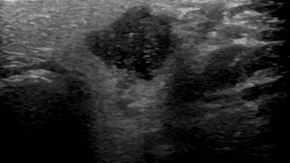
A 66 year old male patient presented with an incidental lesion in the left parotid gland. The patient was asymptomatic and fit and well (See ultrasound image below). Ultrasound and MRI scans showed a well defined lesion in the left parotid gland (1.4 x 1.2 cm) at the junction of the superficial and deep lobe. A provisional diagnosis of pleomorphic adenoma was made.
A fine needle aspirate was obtained from the lesion and sent for histological examination.
Scroll to the right for details and diagnosis.
Whole Slide Histology Image (H&E)
COTM JUNE 2020
Case from Dr Ali Khurram (Sheffield, UK)
CLINICAL DETAILS
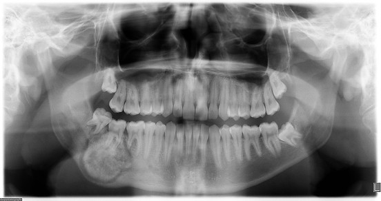
A 25 year old male patient undergoing orthodontic treattment presented with an incidental finding involving the left anterior maxilla. An orthopantomograph (OPG) showed a small radiolucency distal to the upper left vital canine with some evidence of resorption of adjacent teeth (See image below).

The lesion was removed and sent for histological examination.
(click on link below). Scroll to the right for details and diagnosis.
Whole Slide Histology Image (H&E)
COTM MAY 2020
Case from Dr Oluyori Adegun and Dr Reshma Agarwal (UCL, UK)
CLINICAL DETAILS
A 12 year old boy presented with retained ULA and ULC and unerupted UL123. Orthopantomograph (OPG) showed grossly malformed and unerupted teeth in the upper left anterior quadrant. Cone beam CT of the maxilla showed ghost-like outline of the unerupted UL1 and UL3 with enlarged pulp chambers and root canals and a peripheral shell of enamel and dentine (See images below).


The UL123 and associated bone were removed and sent for histological examination.
(click on links below). Scroll to the right for details and diagnosis.
Whole Slide Histology Image 1 Whole Slide Histology Image 2
COTM APRIL 2020
Case from Dr Ali Khurram (Sheffield, UK)
CLINICAL DETAILS
A 17 year old male patient presented with a 28 mm well defined mixed dentisty lesion involving the left body of the mandible in close association with the roots of the LR7 (2nd molar tooth). There was expansion of the right mandible with perforation of the lingual cortex and deviation of the ID canal.
(See image below).
The lesion was biopsied and sent for histological examination.(click on link below).
Scroll to the right for details and diagnosis.
COTM MARCH 2020
Case from Dr Oluyori Adegun and Dr Amrita Jay (UCL, UK)
CLINICAL DETAILS
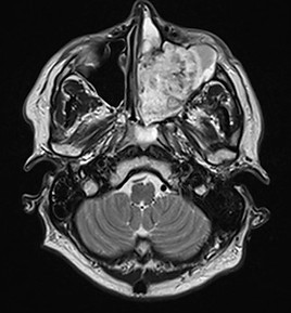
A 50-year-old man presented with left nasal blockage, blurred vision and a feeling of asymmetry of his globe position. He has a past medical history of renal cell carcinoma, secondary polycythaemia and hypertension. MRI scan showed a tumour filling the left maxillary antrum, bulging into the left nasal cavity and extending onto the maxillary alveolus. The left orbital floor was elevated causing mild proptosis (not shown). (See image below).
The lesion was biopsied and sent for histological examination.(click on link below).
Scroll to the right for details and diagnosis.
COTM JANUARY/FEBRUARY 2020
Case from Dr Ali Khurram (University of Sheffield, UK).
CLINICAL DETAILS
A 12 year old boy presented with hyperplastic gingivae in the upper midline regin betwee nthe central incisors. Examination showed a slight swelling of the upper lip in addition to 'cobblestoning' of the buccal mucosa.


An incisional biopsy of the gingival tissue was sent for histopathological examination
(click on link below).
Scroll to the right for details and diagnosis.
Virtual Microscopy/Whole Slide Image